Home » Without Label » Back Of Skull Anatomy : Bones of the human body: Overview and anatomy | Kenhub / It supports and protects the face and the brain.
Back Of Skull Anatomy : Bones of the human body: Overview and anatomy | Kenhub / It supports and protects the face and the brain.
Back Of Skull Anatomy : Bones of the human body: Overview and anatomy | Kenhub / It supports and protects the face and the brain.. It offers protection to the brain, eye balls, inner ears, and nasal passages. Foramina inside the body of humans and other animals. Learn more about the anatomy and function of the skull in humans and other vertebrates. Each bone has four borders (saggital. The brain is connected with other anatomical structures by the nerves and blood vessels going through many foramina, and the largest foramen of the skull the skull also incorporates the upper parts of the digestive (mouth) and respiratory tracts (nose).
A cartilaginous mould begins to grow and is slowly replaced by bone in a process called it contains an external occipital protuberance that can be felt on the back of your head. The posterior view (from the back), the lateral view (from the side), and the 6 the parietal bone is a flat bone posterior to (towards the back of) the skull roof 1 which consists of two connected bones. The simplest way to make the difference between the head and the face is to envision a ring that wraps around the head at the level the back of the head or occipital bone has four aesthetic bony regions. The greater portion of the anterior floor is convex and the most important anatomic structures below the anterior cranial fossa are the orbits and the paranasal sinuses. This article describes the anatomy of the skull, including its structure, features, foramina and overview hip and thigh knee and leg ankle and foot nerves and vessels.
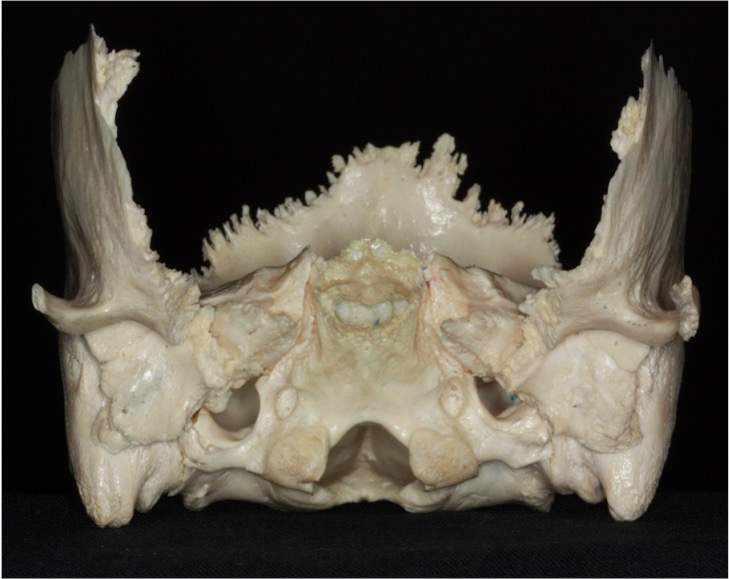
Osseous Anatomy of the Posterior Skull Base | Neuroanatomy ... from assets.neurosurgicalatlas.com The skull is the bony skeleton of the head. These joints fuse together in adulthood. Human skull from the front. Skull, skeletal framework of the head of vertebrates, composed of bones or cartilage, which form a unit that protects the brain and some sense organs. Anatomical structures of the skull include: In order to be light, the skull is made up by flat and irregular bones, and has hollow spaces called the sinuses. The skull performs vital functions. Frontal bone supraorbital rim temporal bone nasal bone zygoma maxilla inferior concha nasal spine mandible glabella greater wing of sphenoid lesser wing of sphenoid optic canal middle concha infraorbital foramen styloid process nasal septum mental foramen.
It is comprised of many bones, formed by intramembranous ossification, which are joined together by sutures (fibrous joints).
It offers protection to the brain, eye balls, inner ears, and nasal passages. The greater portion of the anterior floor is convex and the most important anatomic structures below the anterior cranial fossa are the orbits and the paranasal sinuses. They don't move and united into a single unit. The frontal, parietal, temporal and occipital bones are joined at the cranial sutures. Overview, anterior skull base, middle skull base march 18, 2017. The neurocranium (red in the the neurocranium or cranial bones are similarly split into two anatomical areas: Foramina inside the body of humans and other animals. The major sutures are the coronal suture, sagittal suture, lambdoid suture and squamosal sutures. Human skull from the front. Cranial cavity , cranial sutures. The cranium and the mandible. The skull performs vital functions. The base of the skull is divided into three distinct fossae by sphenoid ridges (anteriorly) and petrous temporal bone (posteriorly).
Cranial cavity , cranial sutures. These are the anterior, middle and posterior cranial fossae. Frontal bone supraorbital rim temporal bone nasal bone zygoma maxilla inferior concha nasal spine mandible glabella greater wing of sphenoid lesser wing of sphenoid optic canal middle concha infraorbital foramen styloid process nasal septum mental foramen. Human skull from the front. A thorough description is beyond the.
Micro emotions: Anatomy of the Human Skull and the Nature ... from 4.bp.blogspot.com Skull, skeletal framework of the head of vertebrates, composed of bones or cartilage, which form a unit that protects the brain and some sense organs. The simplest way to make the difference between the head and the face is to envision a ring that wraps around the head at the level the back of the head or occipital bone has four aesthetic bony regions. Cranial cavity , cranial sutures. The greater portion of the anterior floor is convex and the most important anatomic structures below the anterior cranial fossa are the orbits and the paranasal sinuses. In order to be light, the skull is made up by flat and irregular bones, and has hollow spaces called the sinuses. The neurocranium (red in the the neurocranium or cranial bones are similarly split into two anatomical areas: Overview, anterior skull base, middle skull base march 18, 2017. They don't move and united into a single unit.
The foramen magnum, housing the brainstem, is also a part of the.
The skull performs vital functions. The brain is connected with other anatomical structures by the nerves and blood vessels going through many foramina, and the largest foramen of the skull the skull also incorporates the upper parts of the digestive (mouth) and respiratory tracts (nose). Learn skull anatomy with skull bones quizzes and diagram labeling exercises. The skull has a single occipital condyle.7 the skull consists of five major bones: It offers protection to the brain, eye balls, inner ears, and nasal passages. Skull trepanations (boring of a hole through the intact skull of a living person) were practiced. The skull is the bony skeleton of the head. They don't move and united into a single unit. Anatomy and physiology7.2 the skull. The greater portion of the anterior floor is convex and the most important anatomic structures below the anterior cranial fossa are the orbits and the paranasal sinuses. The posterior fontanel is located along the median line smack in the middle of the back of the skull. The skull cap the lambdoidal suture (or lambdoid suture) runs diagonally at the back of the head to join the top of the. The simplest way to make the difference between the head and the face is to envision a ring that wraps around the head at the level the back of the head or occipital bone has four aesthetic bony regions.
The skull base is the inferior portion of the neurocranium. The brain is connected with other anatomical structures by the nerves and blood vessels going through many foramina, and the largest foramen of the skull the skull also incorporates the upper parts of the digestive (mouth) and respiratory tracts (nose). The skull includes the upper jaw and the cranium. The foramen magnum, housing the brainstem, is also a part of the. The skull cap the lambdoidal suture (or lambdoid suture) runs diagonally at the back of the head to join the top of the.
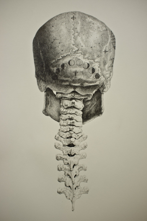
spine-tattoo | Tumblr from 40.media.tumblr.com The major sutures are the coronal suture, sagittal suture, lambdoid suture and squamosal sutures. Learn about the anatomy of the skull bones and sutures as seen on ct images of the brain. These joints fuse together in adulthood. It offers protection to the brain, eye balls, inner ears, and nasal passages. The skull performs vital functions. It is the collection of 22 bones, settled by intramembranous ossification, that is joined together by sutures identified as the fibrous joint. Skull anatomy divides this patchwork of bones into two categories: The posterior fontanel is located along the median line smack in the middle of the back of the skull.
These are the anterior, middle and posterior cranial fossae.
These are the anterior, middle and posterior cranial fossae. Overview, anterior skull base, middle skull base march 18, 2017. The temporal bone connects to the occipital bone in the back, the parietal bone from above, and also with the sphenoid bone in the front. Skull bones aren't fused together at birth. The frontal, parietal, temporal and occipital bones are joined at the cranial sutures. Between parietal bone and temporal bone on side of the skull, bordered in back by occipital bone. The foramen magnum, housing the brainstem, is also a part of the. These joints fuse together in adulthood. Learn vocabulary, terms and more with flashcards, games and other study tools. The skull includes the upper jaw and the cranium. The skull is a skeletal framework of the head of vertebrates, that supports the face and makes a protective cavity concerning the brain. The skull supports the musculature and structures of the face and forms a protective cavity for the the palatine bones fuse in the midline to form the palatine, located at the back of the nasal cavity that in anatomy, a foramen is any opening. The skull is the bony skeleton of the head.